We did it! After nearly three years of work (the initial commit to the code was done on may 24th 2014), the work on the role of apoptosis in the formation of folds in epithelium is out!
I’m very proud for this work. Of course most of the credit goes to Magali Suzanne team. This is top quality biology, and it was really nice being part of the research process. The model itself relies on the marvelous scientific python ecosystem and its supporting community. Special thanks to Tiago for providing the graph-tool library.
The code is described in details in a series of Ipython Notebook that you can read here.
Now for a brief summary on what we did:
[edit 01/23] there’s a nice news and views here where Claudia Vasquez and Adam Martin review the biology and rise interesting questions.
Apoptotic cells last stand - and its consequences for morphogenesis
We showed that, far from being passively eliminated, apoptotic cells do actively influence their environment by increasing the surrounding tissue tension. Indeed, before they die, apopotic cells exert a force that transiently deform the apical surface of the epithelium. This force is then transmitted to the neighbouring cells through an increase in tension which will in turn provoke a change in tissue shape.
Apoptosis is known for its role in morphogenesis, and more specifically in the formation of folds in various developmental contexts; yet the molecular mechanisms implied in those processes remain largely unknown. The formation of folds within an epithelium allows to pass from a bidimentional to a tridimentional tissue and is thus a key step in morphogenesis. Here we showed a new mechanism of fold formation in the Drosophila leg disk. This mechanism not only implies the elimination of apoptotic cells from the tissue, but also their active participation to the tissue remodelling. Indeed, each apoptotic cells within the epithelium generates before it dies a force relying on the establishment of an apico-basal acto-myosin cable. This previously unknown cable drives a transitory deformation of the apical surface of the epithelium, which in turn drives a stabilization of myosin II in the neighbouring cells at the adherent junctions level, as well as an increase in tissue tension. We also showed that the synergistic contribution of several apoptotic cells was necessary to create a myosin II stabilization in the whole neighbouring tissue, a global increase in tension, cells apical constriction and eventually the fold formation.
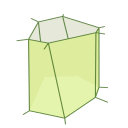
Finally, in order to test whether those apoptotic forces are indeed the initial signal responsible for the change in tissue shape, we devised a 3D model of the leg disk epithelium, based on the pre-existing vertex model published by Farhadifar et al. in 2007. In this bio-mechanical model, we were able to show that apoptotic forces (the apical-basal force, followed by the apical propagation) are both necessary and sufficient to drive fold formation, suggesting that this new mechanism could happen in any type of epithelium.
This work is an important step in the field of morphogenesis and brings up a new dogma on the active role of apoptosis in apoptotic cells.
Bellow is a movie of the formation of the leg joint in vivo (that’s confocal microscopy, apoptotic cells are marked red:
Apical vue of the fold formation on a drosophila leg disk from glyg on Vimeo.
And a movie of the simulated tissue undergoing fold formation:
Fold formation model from glyg on Vimeo.
Of course my work with Magali’s team continues, and there’s a lot more to investigate: the precise mechanism by which the apical-basal force is translated in an apical myosin accumulation, the role of cell polarity in the process, or the role of the peripodial membrane in shaping the tissue, for example. Exciting times!

Comments
comments powered by Disqus